Needle form calcite ("Lublinite") XRDs as calcite, however a casual glance reminds one of aragonite as it takes an acicular form. It is usually associated with soil bacteria, however it has "popped up" in an interesting form: as ribbon helictites. See "Cataloguing Helictites and other capillary-controlled speleothems".
| With a suitable polarising filter, under an optical microscope the needle fibres may be seen. The sketch here gives a general conceptual view of one rod of needle-form calcite. It is comprised of calcite, where each calcite rhomb is attached en echelon to the neighbouring rhomb. |

|
Calcite does not normally precipitate in this form with inorganic processes, however is is a fairly common form when associated with microorganisms (eg Philips and Self, 1987, and also Moore and Sullivan).
Philips and Self (1987) showed several forms of lublinite, some including an organic coating. Possibly organic material holds the shape of the laths, preventing them from coalescing.
Needle Form Calcite in Wollondilly Cave
As part of the cave aragonite project (Rowling, 2004b), I found needle form calcite in fluffy material from "The Loft", Wollondilly Cave (Wombeyan Caves, NSW). The fluffy material also included sediment grains. The area appeared to have had a bat guano deposit a long time ago. The needle fibres were easily visible with a microscope, and the whole material resembled a thatch.I prepared the samples for viewing with an electron microscope, a Phillips SEM 505 at the University of Sydney Electron Microscope Unit. The results are shown below.
Details of the setup are in Rowling, 2004b.
I examined the material with XRD and grouped into major,
minor and trace according to the apparent quantity of material
present in the sample. In the list below, the "major" and "minor" mineral
constituents are reasonably accurate; the trace components however should
be taken with a grain of salt as there may be other substances which
exhibit similar spectra. It would be better to
further test the material (e.g. with mass spectrometry) before one could say
for sure that the trace minerals were actually present.
XRD scans took about 20 minutes per specimen, and subsequent spectral analysis
took about 20 to 30 minutes per specimen, using
the software.
The instruments used were a Siemens Kristalloflex 710D X-Ray generator
and D5000 Diffractometer, at the Electron Microscope Unit at
the University of Sydney.
Details are in Rowling, 2004b.
| Sample ID W144/5 | Minerals detected using XRD | ||
|---|---|---|---|
| This was the most fluffy white material with the longest fibres. Possibly the aragonite is derived from vaterite. | |||
| Suggested mineral origin | Major | Minor | Trace |
| Cave | calcite | aragonite | alunogen |
| ardealite | sacrofanite | ||
| cacoxenite | |||
| diadochite | |||
| manganocalcite | |||
| vaterite | |||
| Detrital | quartz | ajoite | |
| anorthite | boggsite | ||
| biotite-2M1 | colusite | ||
| laumontite | |||
| sherwoodite | |||
| Sample ID W144/11 | Minerals detected using XRD | ||
|---|---|---|---|
| This was the more yellow material with the shorter fibres. | |||
| Suggested mineral origin | Major | Minor | Trace |
| Cave | calcite | ardealite | aragonite |
| cacoxenite | corrensite | ||
| vaterite | |||
| Detrital | quartz | ajoite | |
| boggsite | |||
| colusite | |||
| germanite | |||
| lovdarite | |||
| sherwoodite | |||
| Sample ID W144/12 | Minerals detected using XRD | ||
|---|---|---|---|
| This was the gravel substrate to the fluffy material. | |||
| Suggested mineral origin | Major | Minor | Trace |
| Cave | calcite | aragonite | magnesian calcite |
| heneuite | sodian meionite | ||
| hydromagnesite | earlandite | ||
| lansfordite | liottite | ||
| manganoan calcite | tilleyite | ||
| nesquehonite | skawtite | ||
| baylissite | |||
| defernite | |||
| dypingite | |||
| giorgiosite | |||
| girvasite | |||
| harkerite | |||
| kimuraite-(Y) | |||
| latiumite | |||
| marialite | |||
| sahamalite-(Ce) | |||
| sclarite | |||
| shelkovite | |||
| szymanskiite | |||
| tuscanite | |||
| bradleyite | |||
| sakhaite | |||
| borcarite | |||
| canavesite | |||
| coalingite | |||
| carletonite | |||
| kainosite-(Y) | |||
| callaghanite | |||
| Detrital | quartz | aerinite | |
| high quartz | |||
Some of these may be formed by the action of bat guano on sediments, and others may be derived from reactions with the showcave wire netting. Some minerals are hydrated forms of more common cave minerals. The detrital material appears to be derived from the surrounding volcanics.
The white fluffy material (W144/5 and W144/10) is mainly composed of needle fibre rods, about 1 x 2 μm thick and from 10 μm up to about 1 or 2 mm long. The rods are rectangular with chamfered or 90 ˆ terminations. The material also contains irregular blobs about 2 to 5 μm diameter. The yellow and pale orange material is a mixture of shorter, wider needle fibre rods, along with quartz and other crystal grains and irregular shapes. The yellow material has more needle fibre calcite than the pale orange material which has a higher quantity of quartz and clays. Terminations to the needle fibres are either rectangular or pointed. Some quartz crystal grains are coated in the short fibres.
Some of the minerals listed in the "Minor" headings above may be missing from the various official listings of cave minerals (at least when I did the analyses in 2002). They may have been added since then. Generally a publication is required before a mineral is accepted as a cave mineral.
Electron Micrographs of Wollondilly Cave Needle Form Calcite
I was able to get some detailed pictures of some of my samples using the electron microscope (the old analog SEM 505 was a delight to use). All these pictures are from two tiny samples taken from "The Loft" in Wollondilly cave.
| White fluffy material: sample W144/10. | |
|---|---|
100kb jpg image
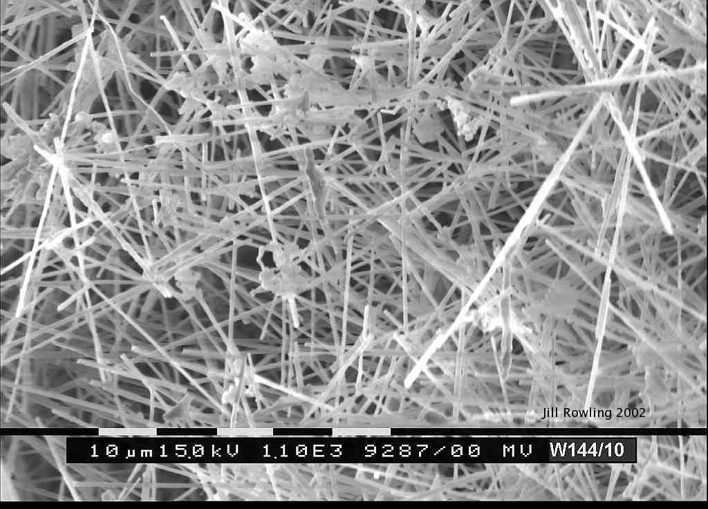
|
The white fluffy needle form calcite resembles a thatch. Small blobs on the end may be a mixture of organics and other material, similar to the XRD listing above for nearby sample W144/5. |
60kb jpg image
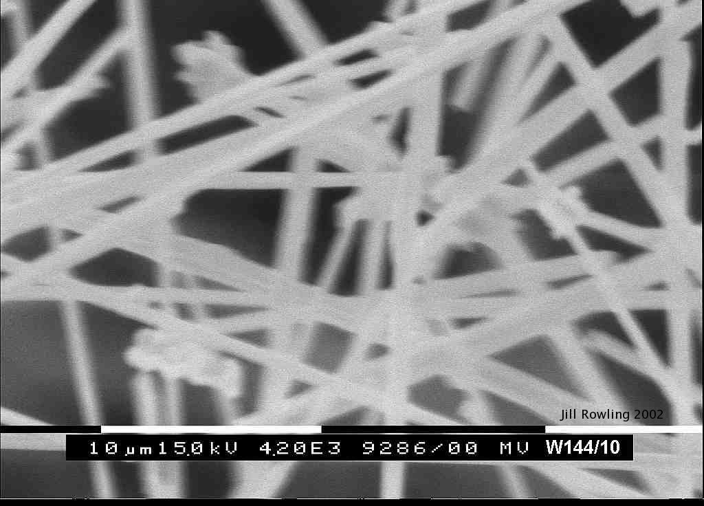
|
Looking closer, the material is very light, with plenty of space between the laths. |
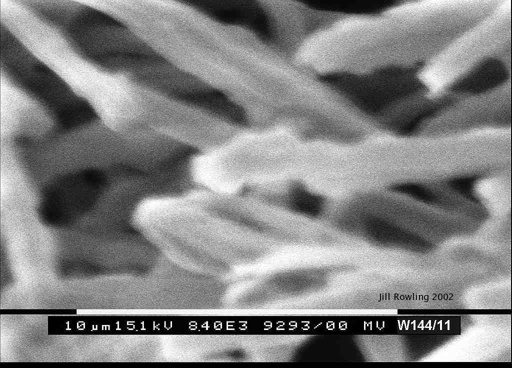
| Yellow fluffy material: sample W144/11. | |
|---|---|
121kb jpg image
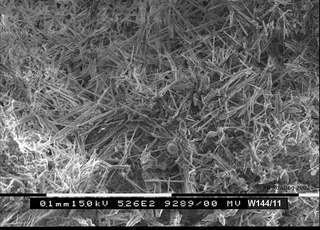
|
Sample W144/11 piece 2 on disc: yellow fluffy material is more dense and has shorter fibres than the white material. |
89kb jpg image
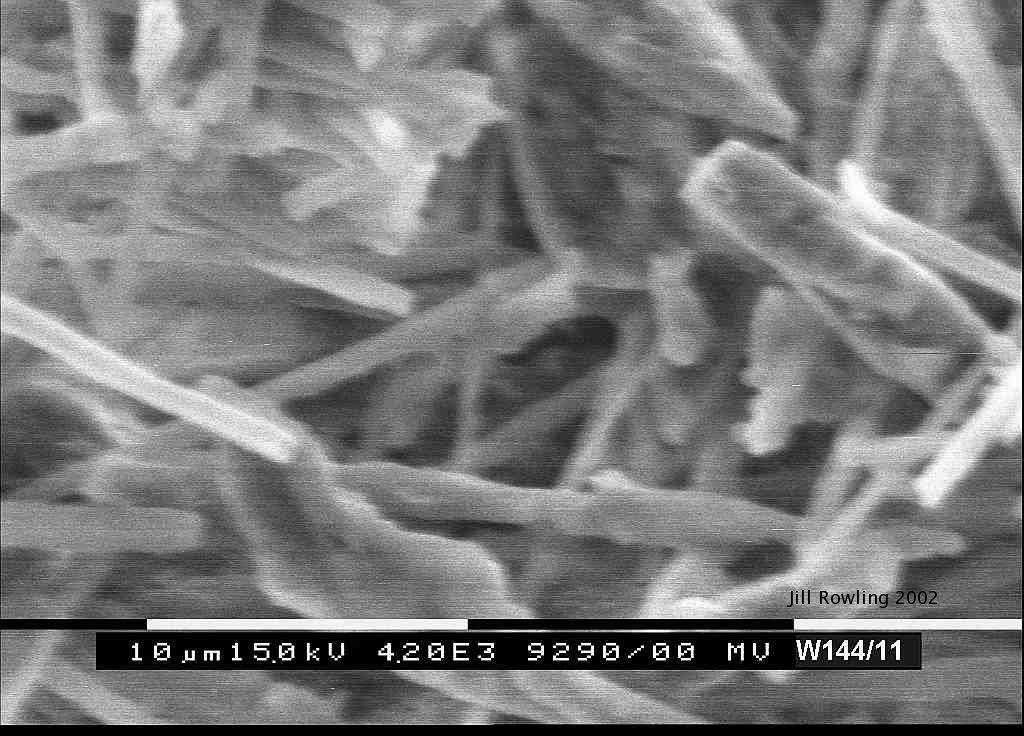
|
Close-in view of sample W144/11, piece 2 on disc, showing termination types of short fibres. Although there are some long fibres, many fibres are not straight; some are bent and joined with others. This thatch looks more "glued together". Some terminations are square (like W144/10) whereas others are pointed. |
122kb jpg image

|
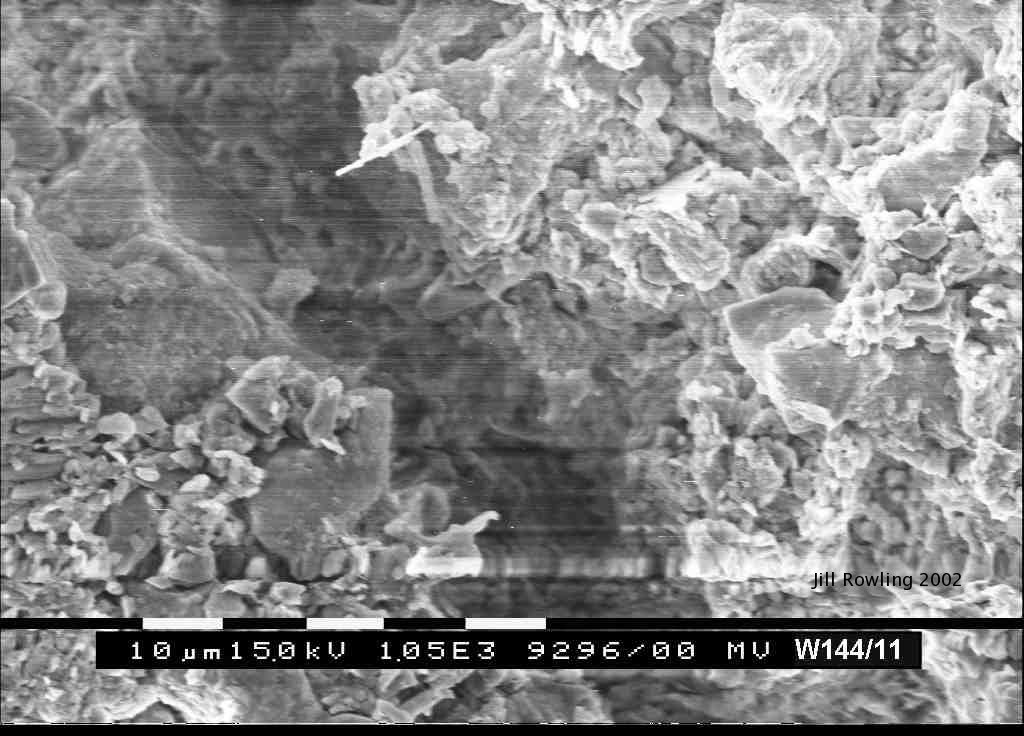
Looking a bit closer at the thatch of sample W144/11, piece 3 on disc. This shows the shorter needle fibres and a spheroidal aggregate (possibly a quartz grain). with some fibres apparently "glued" to others. |
109kb jpg image
|
Close-in view of sample showing termination types of short fibres. Some fibres resemble those in Folk, Chafetz & Tiezzi, 1985 (from bacterial shrubs). Some terminations are square (like W144/10) whereas others are pointed. The ones in the picture vaguely resemble en-echelon calcite. Diameter of laths varies from about 1 μm to 2 μm. Some of these fibres vaguely resemble en-echelon calcite. |
90kb jpg image
|
This is a closer view of the substrate for the "yellow fluffy stuff". The larger grains are probably quartz and there may be clays, mica, feldspar etc. One needle fibre is visible. There is a lot of charging here, possibly from a lack of conductive coating on areas of clay. |
Needle Form Calcite and Ribbon Helictites
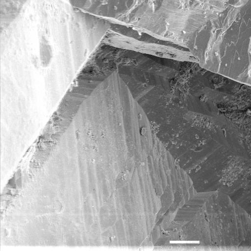
| Surface of ribbon helictite exhibits laths |
|---|
|
Lublinite-like laths are visible with the electron microscope.
The picture is a portion of the surface of a ribbon helictite.
Scale bar is 0,1 mm.
A cropped version of this photo appeared in Rowling, J. (1998) "Ribbon Helictites: A New Category" Helictite 36(1) 2-10. This electron micrograph and sample preparation is by Terry Furey-Grieg, University of Technology, Sydney. |
|
Cosmetic update September 2025. Content created 4th February 2001 and updated 10th September 2025.